NJ hospital upgrades heart procedure to save more lives
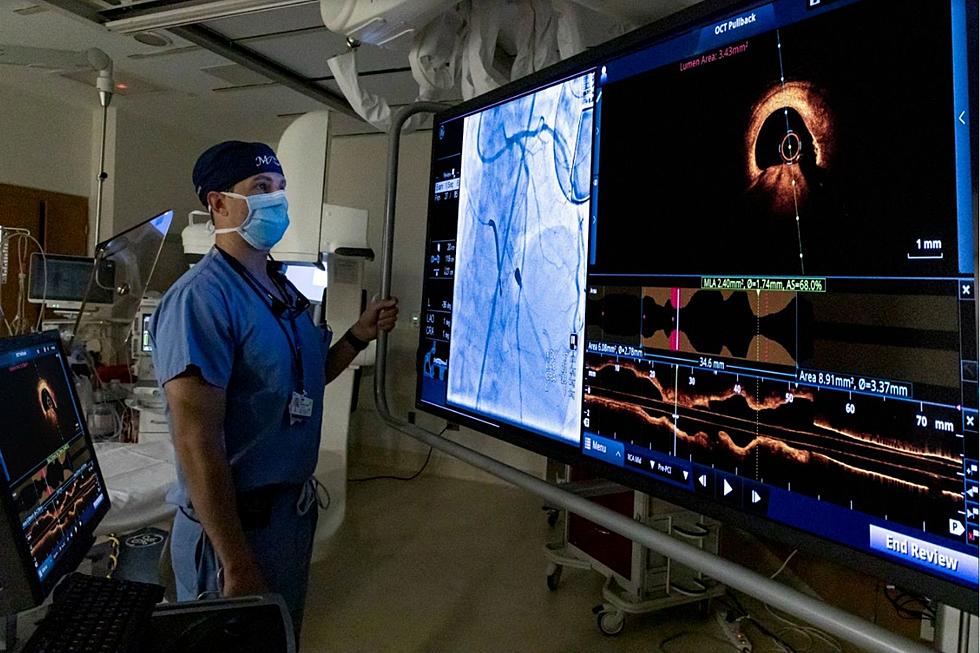
NEPTUNE — Leading-edge technology up and running at Hackensack Meridian Jersey Shore University Medical Center is meant to give doctors a complete picture of what's going on inside patients' coronary arteries, in real time, during procedures that target coronary artery disease.
With the new imaging tool, Optical Coherence Topography (OCT), cardiovascular experts are delivered high-definition images of the interior of the artery by inserting a catheter with a tiny camera. OCT is used during procedures where cardiologists open clogged heart arteries with a stent to improve blood flow to to the heart.

The procedure works to reduce or eliminate patients' symptoms of chest pain and shortness of breath, and helps prevent a heart attack. The hospital has already used the OCT approach on hundreds of patients.
Before now, according to Dr. Matthew Saybolt, director of the hospital's Structural Heart Disease Program, images of the arteries only came from x-rays.
"It's black and white, it's two-dimensional, and this is a three-dimensional problem," Saybolt said. "There are blockages that are hardened with calcium or bony build-up, blockages that have funny angles, that can be smaller or bigger than what we're made to believe on the x-ray."
With the introduction of OCT, Saybolt said, doctors have the ability to overlay the newer 3D images with x-ray photos, giving them the ability to view both the outside and inside of the arteries.
"Patients deserve and want perfection, and this gives us the ability to achieve better results and really provide more personalized treatment options," Saybolt said.
OCT provides accurate measurements inside arteries helping doctors to identify the nature of blockages and reveal lesions, and to help guide stent selection and placement.
"We don't need to make approximations anymore, we can measure what size stent to put in, width and length, and how best to treat any vessel damage," added Dr. Daniel Kiss.
First Responders Appreciation
More From 92.7 WOBM